Gynae & Obstetric
ASS-1701
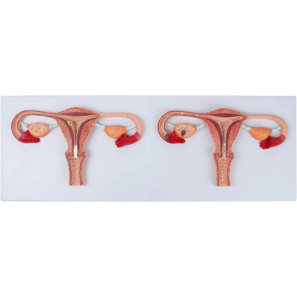
ASS-1701
Fertilization Process Simulator
- Size: 90 x 30x 6.5 cm
- Made of HSP resin
- Two frontal sections of female reproductive organs showing
fertilization process. - Mounted on sturdy wooden frame
- 23 features marked.
- Key card/Manual provided.
Why Choose US
We provide high-quality nursing training models—from human torso replicas to advanced simulators—designed for realistic and effective learning. With trusted quality, affordable pricing, and dedicated support, we help educators and students build the future of healthcare with confidence.
Britannica explains the biological process of fertilization in a clear and academic manner, supporting anatomy and biology education.
Premium Human Fertilization Model for Medical – Detailed Female Reproductive System Teaching Aid
The knowledge of the female reproductive system is essential to students, health care workers, educators in the fields of anatomy, gynaecology, obstetrics, and medical science. Our premium human fertilization model of medical provides an anatomically correct, high-quality graphic and feel portrayal of the complex mechanisms, which take place in the female reproductive system and particularly in the fertilization and initial stages of development. The female reproductive system anatomy is also greatly applicable in both academic and clinical settings using this model.
Table of Contents
What Is the Female Reproductive System?
The female reproductive system is a complex system of organs that produce hormones, ovulate, conceive, carry a pregnancy to term and give birth. It encompasses external and internal structures like vagina, uterus, fallopian tubes, ovaries and related body parts. The different parts do work together to have a particular role in reproduction such as the housing and release of the ova (eggs) and the subsequent support of an embryo once fertilized. All these structures are a representation of female reproductive system organs, which are vital in human reproduction.
The internal organs are also well-organized to allow the transport of gametes and foster fertilization whereas the outer structures safeguard these systems and support reproductive functions. This system coordinates hormonal regulation, menstrual cycles, complex processes in the body and so on.
Why This Model Matters
Textbooks may not suffice to understand anatomy and physiology. Our female reproductive system model fills in the gap between theory and the real world:
Three-Dimensional Visualization: This model contrasting the flat images brings the female reproductive system alive and present and it is detailed with the sections of the front part vividly present in comparison to a typical system of the female reproductive system diagram.
Teaching Aid: It would be a perfect tool in the classroom, laboratory, and medical training centers where instructors can show the process of fertilization in an easy and accurate way.
Practical Learning: The students are able to learn about the spatial relations between organisms such as the ovaries, fallopian tubes, uterus, and so on.
Durable Build: The model is made of high-quality HSP resin and fixed on the strong wooden frame which makes it long-lasting and can be used several times.
Key Features of the Model
High-Definition Anatomical Sections: Two frontal sections are present with the key internal organs of the female reproductive system, in particular, the ovary, fallopian tube, and uterus.
Knowing the Fertilization: Assists in showing the way an egg that has been released by the ovary moves through the fallopian tubes and joins sperm, which is the most important point of fertilization in the female reproductive system.
Durability: HSP resin of professional quality was used to make a strong construction that is realistic and dependable as an educational tool.
Mounted Display: The wooden stand has a stable and appealing foundation to be stood on and be displayed in learning settings.
Realistic Detailing: Colours and textures are made to resemble the real anatomy so as to be as educational as possible.
Learn the Female Reproductive System in Detail
This instructional format assists in improved understanding of female reproductive system including:
Internal Organs
Ovaries: These are structures which are paired where ova are produced and hormones like estrogen and progesterone are released.
Fallopian Tubes: These are the tubes that hold the egg once ovulation has taken place and is where a fertilized egg is usually fertilized.
Uterus: It refers to a muscular organ on which the fertilized egg implants and grows to become a fetus.
Cervix and Vagina: Lower tracts in which sperm penetration and delivery of a child takes place.
Knowledge about the location, function and interrelationships of these parts supplements’ prior knowledge of reproduction in humans and contributes to the explanation of such cycles as menstruation, conception and early pregnancy.
Who Should Use This Model?
This educational aid would suit:
- Nursing and Medical Students
- Anatomy & Physiology Classes
- Gynecology and Obstetrics Training
- Health Education Workshops
- Clinics and Hospitals
This explicit display of the female reproductive system enables the learner to have a graphical view of the process of fertilization that cannot be realized solely by textbooks and thus a better learning and comprehension of complex biological processes.
Increase Learning Through Experience
With this female reproductive system model, educators are able to facilitate interactive activities that will promote discussion, exploration and critical thinking. Students are able to follow the path of the egg, learn about each organ, and connect with actual reproduction ideas in the real world at the same time- the student has academic and clinical confidence.
This is an anatomical resource that cannot be ignored to make your medical education more comprehensive and effective.
Why Choose Us
Our educational models are anatomically correct and reliable and are meant to be used in real life. The durability, clarity and professional craftsmanship are part of our products. Our priorities are the quality, price, and customer service. We offer reliable resources to teachers, learners, and medical workers with our industrial knowledge to transform teaching, learning, and prolonged educational achievement.
FAQs
Q1: What does this model illustrate?
It approaches the process of fertilization and the functioning of the female reproductive system in a clear and easily understandable and practical manner.
Q2: Does it suit medical students?
Yes, it is perfect for medical, nursing, and anatomy students learning the anatomy of the female reproductive system.
Q3: Are the internal organs detailed in the model?
Yes, it has all major female reproductive system organs displayed in a way that is easier to understand.
Q4: Is this applicable in a classroom teaching?
Definitely, it is a valid substitute to a conventional female reproductive system diagram in classrooms.
Q5: Can the material be used on a long-term basis?
Yes, it is made of durable HSP resin and is intended to be used by professionals on a regular basis.