Learning about the female reproductive system is the necessary concern of students, healthcare-training people, or people who want to know the correct information about human biology. Not only does this system control menstruation, hormones, and reproduction, but it also is crucial to the overall health and development. Proper understanding of structure and the functions will enable one to have a better understanding of fertility, pregnancy, and common medical conditions. This guide describes the organ, procedure and biologic coordination involved in it with explanations that are medically reliable, and the learner centred format that is in tandem with contemporary educational provisions.

Table of Contents
Biological Framework of Reproductive Design
The female reproductive system is a complex network of both internal and external organs which work together to help the ovulation process, fertilization and gestation. Each of these elements has a particular contribution to the production of gametes, the hormonal equilibrium, and fetal growth. This system is dynamic and the reaction to endocrine signals and the environment takes place between puberty and menopause. Due to its interrelation, cycles, fertility, or even general wellbeing can be impacted due to minor disruptions.
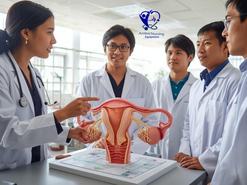
The use of three-dimensional learning is getting more prevalent in medical education as students have the opportunity to visualize the interrelationships between organs. The spatial orientation and physiological interaction are explained with the help of teaching instruments and anatomical models provided by Ambay Nursing Equipment. These resources facilitate proper understanding especially in nursing and allied health training. To find out about the selection of effective models of medical training, which increase the educational effectiveness, you can refer to this blog complete guide to choosing the right CPR manikin.
Organizational Structure and Major Elements
The complete knowledge of female reproductive system anatomy commences with the identification of its fundamental organs. The central reproductive pathway is made up of ovaries, fallopian tubes, uterus, cervix, and vagina which are located internally. Outside the body, the vulva shields openings inside the body and has a role to play in sexual performance. As a combination, these organs comprise the main female reproductive system organs, each with a specific duty.
The ovaries are capable of generating ova and releasing estrogen and progesterone. Released ova are trapped by the fallopian tubes and offer the place of fertilization. Implantation and pregnancy occur in the uterus which is made up of muscular walls and a receptive lining. The cervix controls the movement between uterus and vagina and the vagina is the copulatory canal and additionally the birth passage. These structures when found in the same context show the integrated architecture of the female reproductive system.
Hormonal Control and Cyclical Function
The female reproductive system revolves around hormonal control. The signals are co-ordinated by the hypothalamus and pituitary gland and stimulate the activity of the ovary leading to cyclic variations called the menstrual cycle. The follicular development and thickening of the uterine lining is mediated by estrogen and progesterone prepares the uterus in case implantation occurs.
This is not just the reproductive cycle; it affects the density of the bones, the health of the heart, and the emotional stability. Cycle regularity can change due to any interruption, whether caused by hormonal imbalance, stress or disease. Learning institutions frequently use physical educational similarities, such as a female reproductive system model, to show the endocrine feedback mechanisms and anatomical reactions at every stage.
Visualization by Educational Models
Anatomy is vital in health care training. The female reproductive system diagram is used to simplify the complex relationship of organs by showing them in their proportions and orientation. Diagrams also focus on space organization, and illustrate the relationship between ovaries and fallopian tubes, the position of the uterus relative to the cervix, and the relationship between tissues around the organs and the reproductive processes.
Three dimensional models also contribute to more understanding, as they provide hands-on learning. Ambay Nursing Equipment products are integrated in many of the training centers due to their well-built models that help to support theories with visual representations. Such instruments are especially useful in learning about the differences in uterine shape, position of the cervix, and the form of the pelvis.
Between Ovulation and Conception
Ovulation is the first step in the process of fertilization. A mature egg is discharged by each ovary every month and is pulled into the fallopian tube by cilia. The sperm that is deposited during intercourse enters the cervix and the uterus to land on the tube. When a viable sperm enters into the egg, the process of fertilization takes place leading to the formation of a zygote.
In a few days, the zygote separates and moves to the uterus whereby implantation is carried out in the endometrial lining. This is a sensitive procedure that underscores the accuracy of the female reproductive system. Some small structural impairments or hormonal disproportion can block successful conception, and it is essential to learn about the workings.
Uterine Adaptation and Pregnancy Support
After the implantation process, the uterus is what becomes the center organ of developing the fetus. Its muscular walls stretch as the placenta develops and this process aids in the exchange of nutrients and oxygen. Hormonal changes will inhibit additional ovulation and hold on to a pregnancy. The cervix does not open yet it is sealed, preserving the growing embryo and the vagina prepares to deliver a baby.
Scaled replicas of the anatomy commonly are used in clinical training to demonstrate how the size of the uterus varies with trimester through the use of products sold by the Ambay Nursing Equipment. These studies offer practical insights into maternal adaptation and fetal development, which support the physiological integrity of the female reproductive system.
Basic Disorders and Health Consciousness
The female reproductive system is susceptible to several conditions that include polycystic ovarian syndrome, endometriosis, fibroids, infections, and hormonal disorders despite its efficiency. Quick diagnosis is based on the knowledge of normal anatomy and normal functioning. To illustrate, the presence of irregular cycles can be an indication of endocrine imbalance, whereas chronic pelvic pain can be a sign of structural abnormalities.
Anatomical charts, models, and diagnostic imaging help to identify such issues by health professionals. To enable learners to distinguish between normal and pathological outcomes, education providers usually incorporate a female reproductive system model into the curriculum. Knowledge gives people the strength to obtain medical attention in time and keep their reproductive system healthy.
Ethical Understanding and Clinical Education
In addition to biological facts, there is an ethical sensitivity, patient privacy, and informed consent related to learning about the female reproductive system. Proper anatomical teaching can guarantee a respectful approach by healthcare professionals to patients. Formal education minimizes the myths and encourages evidence-based practice.
The use of hands-on options provided by Ambay Nursing Equipment is becoming more and more common in modern nursing programs to help bridge the gap between theory and practice. Through learning how other professionals present authentic simulations of the female reproductive system anatomy, students gain confidence in clinical evaluation, patient education, and support in the procedure.
Lifespan Perspective
The female reproductive system changes between menarche and menopause. Puberty starts hormonal processes, fertility is maintained throughout the reproductive years, and menopause is the end of the ovulation process. All the stages are associated with the specific physiological features that demand different health care strategies.
Awareness of such transitions assists the professionals in advising patients on birth control, pregnancies, hormone replacement, and precautionary screening. Clarity with easy visualization of diagrams, charts, and other physical replicas will guarantee a uniform understanding of educational levels, which will strengthen long-term reproductive health literacy.

Conclusion
The female reproductive system is one of the most complex biological networks within a human body. It facilitates reproduction through the coordinated anatomy, careful hormonal regulation and adaptation of function, and affects the general wellbeing. Through the investigation of the organs of the female reproductive system, visualization of the pathways using a female reproductive system diagram, and reinforcement of learning using a female reproductive system model, the students and professionals can get practical understanding of fertilization, pregnancy, and health management. Extensive education does not only help in educational achievement, but also ensures informed, respectful, and effective healthcare practices at all stages of life.
FAQs
Q1. What is the major purpose of the female reproductive system?
The female reproductive system performs the functions of egg production, control of vital hormones and fertilization as well as maintenance of pregnancy and childbirth.
Q2. What is the significance of learning about reproductive anatomy in healthcare?
Proper understanding of the reproductive structures enables the medical profession to diagnose the conditions, offer proper treatment and offer proper patient education.
Q3. What is the effect on the learning of human reproduction using visual aids?
Illustrations assist students to learn the position of organs, biological functions and the interactions between various structures, simplifying complex concepts.
Q4. Why do we need physical teaching models when teaching medicine?
Enhanced spatial knowledge, hands-on learning, and long-term retention of anatomical concepts are also found to be improved with the help of three-dimensional teaching aids.
Q5. Is it possible that fertility problems can be observed even when the organs are healthy?
Yes. Even normal looking structures can be affected by hormonal imbalance, inflammatory or microscopic abnormalities that can disrupt reproductive functionality.