Skeleton Models
ASS-102B
ASS-102B
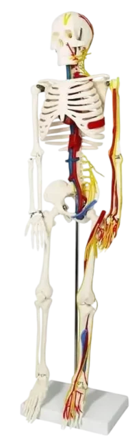
Human Skeleton
- Size 85cm Medium.
- Made of PVC plastic.
- Shows nerves and blood vessels.
- Depicts the position, course and distribution of main arteries and peripheral nerves
- Can be employed as a visual aid in the instruction of anatomy to students of medicine.
Why Choose US
We provide high-quality nursing training models—from human torso replicas to advanced simulators—designed for realistic and effective learning. With trusted quality, affordable pricing, and dedicated support, we help educators and students build the future of healthcare with confidence.
For a detailed understanding of the human skeletal system and its functions, trusted anatomy resources like MedlinePlus and Kenhub provide clear explanations and visual guidance.
Human Skeleton Model for Medical Education and Anatomy Demonstration
Understanding human anatomy is easier when students are able to view and study structures in a real sense. The human skeleton model is created to depict a perfect replica of the skeletal structure; hence it is a good learning tool in medical institutions, nursing colleges, physiotherapy centers and even in science laboratories. We have provided a model that provides hands-on learning and enables students to have a solid background in anatomy by combining anatomical accuracy with robust construction.
Our models are designed to imitate the natural framework of bones and joints in a way that the learners will understand how the skeletal system supports the human body. By working with our human anatomy skeleton model directly, the students are in a better position to visualise the location of the bones, the motions of the joints, and the anatomical associations, better than with textbooks alone. The given practical form of learning enhances perception and interaction in classroom demonstrations and laboratory sessions.
Table of Contents
Realistic Design of Detailed Anatomy Study
Each human skeleton teaching model is formed in life-size proportions so that learners can research the skeletal structure the way it is in the human body. The model has detailed bones, well-defined joints and precise anatomical landmarks, which can assist teachers to explain complex concepts in a better way.
The key features of the skull are also drawn with care such as a detachable skull cap and mobile mandible that enable the students to observe the structures of the skull and the jaw movement. Single teeth are also fitted to illustrate the anatomy of the skull and alignment of the teeth.
The full body human skeleton model is also a representative of other important body parts of the human body like the cervical area, the spinal column, and the sacral canal. These aspects enable students to learn about the structural alignment of the spine and the human skeletal system.
Durable Construction for Long-Term Use
Our human anatomy skeleton model is produced with high-quality lightweight PVC which is durable. This enables the model to be tolerant of repetitive use in demonstrations and rehearsal sessions.
Since the material is washable and cannot be damaged easily, the model can be used in the busy training setups where trainees are constantly exposed to the model.
Simple Demonstration with Adaptable Structure
Our human skeleton model with stand is designed to accomplish the task of demonstrations in the classroom more conveniently. The skeleton is placed on a free-standing table that supports the structure and makes it visible when lecturing. The stand also has smooth rollers so that instructors can simply transfer the model between classrooms or demonstration spots.
Some sections of the skeleton can be removed and this enables the educator to isolate certain bones and explain anatomical structures in a more understandable way.
The Main Characteristics of Our Skeleton Model
- A full-size human skeleton model to study anatomy correctly
- Extensive human anatomy skeleton model with realistic bone structure
- Skull cap removable and movable mandible, cranial study
- Demonstration and learning of specific limbs by detaching them
- Long-term PVC construction intended to be used in institutions
- A stable human skeleton model with stand to facilitate display and movement easily
- School, medical college, and laboratory ideal human skeleton teaching model
Best in Medical Training and Education
Our full-body human skeleton model has been popular in medical training facilities where precise anatomical modelling is a strong requirement. The skeletal structures can be well depicted by the teachers and the students can also get good hands-on experience, which enhances their knowledge of the human body.
Our models can be used to make classroom learning more interactive by integrating visual learning with a practical observation. Medical, nursing, physiotherapy and biological science students can use the tool that enables them to study the anatomical structures firsthand.
FAQs
It is employed in schools, colleges, and medical training institutions in teaching and studying the structure of the skeletal system of the human body.
Yes, the model has a stable stand where it can be easily displayed and moved when demonstrating in the classroom.
It is appropriate for medical students, teachers, health care trainees and biology students.
It allows the learners to have a clear perception of the bone structure, joints and alignment of the body through visual and physical learning.
Yes, it is constructed of study materials that can withstand constant use in the classrooms and the laboratories.